Treatment of Cerebral AVMs through Cerebral Angiography
Treatment of Cerebral AVMs through Cerebral Angiography
Cerebral arteriovenous malformations (AVMs) are vascular abnormalities that can cause bleeding or damage surrounding tissues. Some people do not experience symptoms until the cerebral AVM starts bleeding. The goal of treating cerebral AVMs through cerebral angiography is to shrink them or stop the blood flow through them.
What are Cerebral Arteriovenous Malformations (AVMs)?
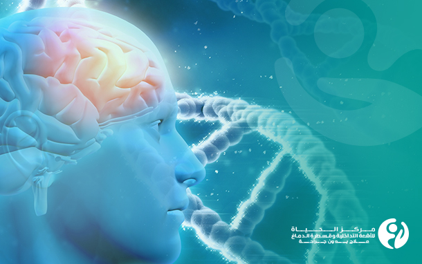
A cerebral AVM is an abnormal tangle of blood vessels caused by the absence of capillaries between the arteries and veins. The tangle consists of arteries that supply blood to the brain and veins that drain blood from the brain tissues.
Blood moves through the body in a regulated, closed circuit of blood vessels. Arteries carry oxygen-rich blood from the heart to the brain and other tissues. Veins return the oxygen-depleted, nutrient-poor, waste-rich blood from the tissues to the heart and lungs.
Normally, the exchange occurs in the capillaries that connect the arteries and veins. In cerebral AVMs, the capillaries between arteries and veins are missing; this allows the high-flow arterial blood to connect directly to the veins, which are not adapted to handle high blood pressure. As a result, the abnormal connection between the artery and vein can cause the blood vessels to rupture and bleed in the brain.
How common are cerebral arteriovenous malformations (AVMs)?
Brain arteriovenous malformations are rare. They occur in less than 1% of people. Anyone can be born with a cerebral arteriovenous malformation.
What causes cerebral arteriovenous malformations?
The causes of arteriovenous malformations are not fully understood, but they develop as the fetus grows (they are congenital), and their occurrence in some families is rare.
What are the symptoms and complications of brain arteriovenous malformations?
• Brain hemorrhage/stroke: A brain AVM causes blood to flow forcefully from the arteries and puts pressure on the veins, which have weak walls and cannot adapt to the high blood flow pressure. If the veins cannot withstand the blood pressure, they may rupture and bleed. Bleeding in the surrounding tissues can cause permanent damage. If the bleeding is severe, it can be fatal.
The bleeding is the greatest risk of a brain AVM and occurs in 38-73% of patients. Bleeding from a brain AVM can cause stroke, brain damage, or seizures.
• Seizures: A surge of electrical activity in the brain may cause fainting and uncontrollable muscle movements.
• Aneurysm: An aneurysm is a balloon-like bulge in the walls of a blood vessel. An aneurysm develops because of a weakening of the blood vessel wall and occurs in about 50% of all brain AVMs. Aneurysms associated with a brain AVM increase the risk of rupture (bleeding) and bleeding-related symptoms.
• Tissue oxygen depletion: Without the capillaries between the arteries and veins, oxygen and nutrients do not reach the tissues containing the brain AVM. The nerve tissue can die, leading to brain damage that can affect thinking, memory, language comprehension, and gradual neurological deficits.
• Coma and death: Especially in cases of major brain hemorrhages.
How are cerebral arteriovenous malformations treated?
The goal of treating arteriovenous malformations (AVMs) of the brain is to reduce or permanently eliminate the risk of bleeding. However, it is difficult for surgery to completely remove these vascular malformations, as the center of the arteriovenous malformation develops new connections to the arteries and can grow back.
Al Hayat Center for Interventional Radiology and Neurointervention in Karbala, Iraq is known for treating brain arteriovenous malformations using the latest global methods, by occluding the arteries with coils or glue materials placed inside the malformation through cerebral catheterization.
Cerebral catheterization:
Cerebral catheterization is very effective in treating cerebral arteriovenous malformations, and it is the first line of treatment, as it blocks or destroys the abnormal arteries while preserving the normal arteries.
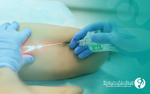
The devices at Al Hayat Center for Interventional Radiology and Neurointervention allow the insertion of a narrow catheter in brain blood vessels through a small incision in the patient's leg. When the catheter reaches the brain, it fills the blood vessel with a metallic thread called coils, which prevents blood from entering the vascular malformations and thus protects the patient from further rupture.
The interventional radiologist at Al Hayat Center for Interventional Radiology and Neurointervention in Karbala uses imaging guidance techniques to navigate the catheter, a small plastic tube, and deliver it to the site of the arteriovenous malformation. Once tthe catheter reaches the arteries feeding the malformation, medical glue or coils are injected to stop blood flow through the AVM, causing it to shrink and close.
These simple interventions can be performed with just light anesthesia. The complete shrinkage of the arteriovenous malformations can take 4-6 weeks.
Frequently asked questions and answers about the treatment of cerebral AVMs via cerebral angiography:
Can arteriovenous malformations be fatal?
Yes, they can be. The severity of a cerebral arteriovenous malformation depends on its size and location. The massive bleeding resulting from the rupture of a cerebral arteriovenous malformation in the brain can be fatal.
Does the location of the head pain indicate the location of the cerebral arteriovenous malformation?
Not necessarily. It may be possible, but in most cases, the location of the headache is not specific to the area of the cerebral arteriovenous malformation.
Do brain hemorrhages from arteriovenous malformations always cause significant brain damage?
No, some cases of very small brain hemorrhages, called microbleeds, cause limited damage and few symptoms. However, over time, multiple microbleeds can increase the risk of developing dementia and impaired cognitive functions.
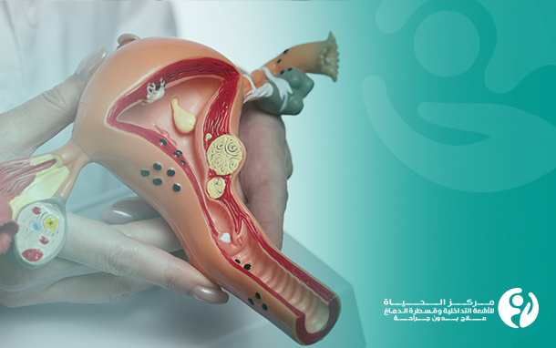
What is the difference between cerebral arteriovenous malformation, aneurysm, cavernous malformation, and arteriovenous fistula?
Aneurysms are weakened and dilated areas in an artery, often forming at branch points. Aneurysms can occur as a complication of cerebral arteriovenous malformations.
Cavernous malformations are a type of vascular disease that differ from cerebral arteriovenous malformations, as they contain slow-moving and usually clotted blood. Cavernous malformations may leak blood but usually do not bleed profusely like cerebral arteriovenous malformations and often do not cause symptoms.
Arteriovenous fistulas are abnormal connections of blood vessels. Arteriovenous fistulas can occur in the tissues covering the brain and spinal cord. Unlike cerebral arteriovenous malformations, which occur within the brain tissue, arteriovenous fistulas are more associated with head trauma or infection.
Al Hayat Center for Interventional Radiology and Neurointervention in Karbala treats all these conditions for the first time in Iraq with great success.